Upper Leg Tendon Anatomy - Muscles Of The Posterior Leg Attachments Actions Teachmeanatomy : Originates from the lateral condyle of the tibia and the medial surface of the fibula.
Upper Leg Tendon Anatomy - Muscles Of The Posterior Leg Attachments Actions Teachmeanatomy : Originates from the lateral condyle of the tibia and the medial surface of the fibula.. Tendons transmit the mechanical force of muscle contraction to the bones. Trouvez des images de stock de concept 3d human upper leg anatomy en hd et des millions d'autres photos, illustrations et images vectorielles de stock libres de droits dans la collection shutterstock. Muscle/tendon inflammation and pain along anterio… The tendons that control movement in your hands, wrists and fingers run through your forearm. By spicer mcleroy in tutorials.
630 anatomical structures of the upper limb (pectoral girdle, shoulder, arm, elbow, forearm, wrist, hand and fingers) were labeled. Tendon, tissue that attaches a muscle to other body parts, usually bones. The tendons of the edl can be palpated on the dorsal surface of the foot. By spicer mcleroy in tutorials. Anatomy of leg and foot human muscular system stock vector.,category:anatomy of the human leg,muscles of the leg and foot classic human anatomy in motion:
The Anatomy Of A Runner It S All About That Bass The Upper Leg Glutes The Fartlek from marciaruns.files.wordpress.com The pads of the machine are situated at the achilles tendon. By spicer mcleroy in tutorials. The artist's guide to the.,muscles that lift the arches of the feet and more. The tendons of the edl can be palpated on the dorsal surface of the foot. What are the functions of patella. Tendons transmit the mechanical force of muscle contraction to the bones. It serves to attach the plantaris, gastrocnemius (calf) and soleus muscles to the calcaneus (heel) bone. Hands are outstretched, holding onto the handles of the bench.
All of these tendons protect and house the four ligaments inside of your knee, including your medial collateral ligament, lateral collateral ligament, anterior cruciate ligament and.
Injuries to the achilles tendon are very serious. Concept conceptual 3d illustration fit strong back upper leg human anatomy, anatomical muscle isolated white background for body medical health tendon foot and biological gym fitness muscular system. Fascia of the upper limb. It is the largest tendon of the parts of leg. Tendons are fibrous cords attached to muscles and bone. Understanding the function and anatomy of the peroneus longus can help you make the best choices for your care if you have suffered and injury there. ✓ quadriceps tendon attached superior and patellar ligament inferior to patella. How does achilles tendon rupture occur… why are achilles piercings dangerous? And it is also critical to the walking process. Tendons are thick bands of tissue that connect muscles to bone. Superficial veins of upper limb , anatomy : Your hamstring tendons run behind your knee and meet your patellar tendon. This mri wrist coronal cross sectional anatomy tool is absolutely free to use.
Lateral (fibular) collateral ligament (fcl) upper part middle part lower part popliteus tendon (pt) upper part i. Tendon, tissue that attaches a muscle to other body parts, usually bones. Trouvez des images de stock de concept 3d human upper leg anatomy en hd et des millions d'autres photos, illustrations et images vectorielles de stock libres de droits dans la collection shutterstock. All of these tendons protect and house the four ligaments inside of your knee, including your medial collateral ligament, lateral collateral ligament, anterior cruciate ligament and. Superficial veins of upper limb , anatomy :
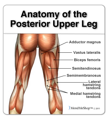
Anatomy Of The Hamstring Upper Leg from aidyourhamstring.com It serves to attach the plantaris, gastrocnemius (calf) and soleus muscles to the calcaneus (heel) bone. Tendons are thick bands of tissue that connect muscles to bone. When a muscle contracts, the tendon pulls on the bone causing the joint to move. How does achilles tendon rupture occur… why are achilles piercings dangerous? Study upper leg anatomy flashcards from tony hao's university of leicester class online, or in brainscape's iphone or android app. Alas, anatomical name changes occur slowly over time and the traditional peroneus name continues to be used. Tendons are fibrous cords attached to muscles and bone. Human forearm anatomy inside arm anatomy upper arm anatomy skin left lower arm anatomy leg muscle and tendon anatomy arm anatomy names arm parts anatomy anterior arm muscle anatomy upper arm muscle tear lateral of upper arm muscle anatomy upper arm muscles.
Alas, anatomical name changes occur slowly over time and the traditional peroneus name continues to be used.
Tendon, tissue that attaches a muscle to other body parts, usually bones. Upper leg anatomy and function. Use the mouse scroll wheel to move the images up and down alternatively use the tiny arrows (>>) on both side of the image to move the images. Superficial veins of upper limb , anatomy : Upper limb trauma programme injuries. Spicermanyt at checkout for 40% off this tutorial! There is no real division between the core and the upper leg; What are the functions of patella. Tendons are fibrous cords attached to muscles and bone. The tendons that control movement in your hands, wrists and fingers run through your forearm. Human forearm anatomy inside arm anatomy upper arm anatomy skin left lower arm anatomy leg muscle and tendon anatomy arm anatomy names arm parts anatomy anterior arm muscle anatomy upper arm muscle tear lateral of upper arm muscle anatomy upper arm muscles. Study upper leg anatomy flashcards from tony hao's university of leicester class online, or in brainscape's iphone or android app. Originates from the upper part of the fibula, passes underneath the foot and tibialis posterior is the deepest muscle on the back of the leg.
Alas, anatomical name changes occur slowly over time and the traditional peroneus name continues to be used. Originates from the lateral condyle of the tibia and the medial surface of the fibula. Hands are outstretched, holding onto the handles of the bench. Upper limb trauma programme injuries. This mri wrist coronal cross sectional anatomy tool is absolutely free to use.
Pin On Health Metabolism from i.pinimg.com .16 penile numbness and perineum tenderness.18 any suggested exercises or stretches?.22 leg musculature 209 elbow tendonitis and saddle sores. By spicer mcleroy in tutorials. Fascia of the upper limb. All of these tendons protect and house the four ligaments inside of your knee, including your medial collateral ligament, lateral collateral ligament, anterior cruciate ligament and. Understanding the function and anatomy of the peroneus longus can help you make the best choices for your care if you have suffered and injury there. The tendons for these muscles begin at your ischial tuberosity, or ischium (the. It is the largest tendon of the parts of leg. Muscle/tendon inflammation and pain along anterio…
There is no real division between the core and the upper leg;
Originates from the upper part of the fibula, passes underneath the foot and tibialis posterior is the deepest muscle on the back of the leg. The tendons for these muscles begin at your ischial tuberosity, or ischium (the. 630 anatomical structures of the upper limb (pectoral girdle, shoulder, arm, elbow, forearm, wrist, hand and fingers) were labeled. Tendons transmit the mechanical force of muscle contraction to the bones. Originates from the lateral condyle of the tibia and the medial surface of the fibula. Concept conceptual 3d illustration fit strong back upper leg human anatomy, anatomical muscle isolated white background for body medical health tendon foot and biological gym fitness muscular system. Lateral (fibular) collateral ligament (fcl) upper part middle part lower part popliteus tendon (pt) upper part i. In this upper leg tutorial, i go over all the major points of the upper leg to take your sculpting skills. Upper leg anatomy and function. Spicermanyt at checkout for 40% off this tutorial! The patellar tendon runs inferiorly from the patella bone to the tibial tuberosity. Collectively, they act to dorsiflex and invert the foot at the ankle joint. Tendons are thick bands of tissue that connect muscles to bone.


0 Komentar